Rhabdocline Needle Cast
Rhabdocline weirii (fungus)
HOST Douglas-fir
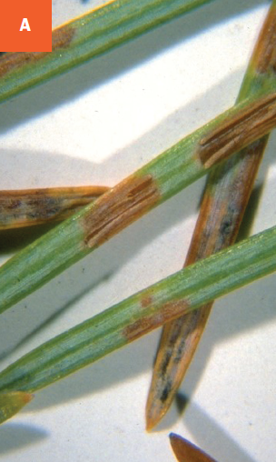
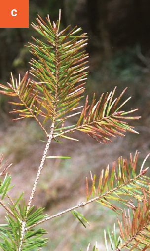
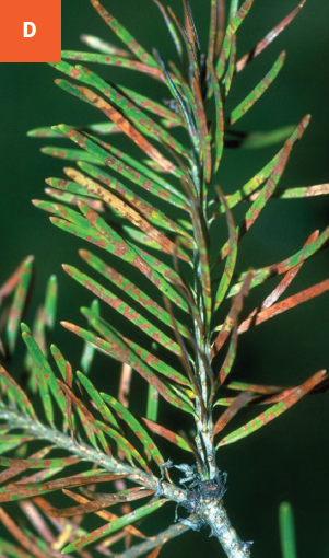
DAMAGE/SYMPTOMS In late summer to fall, yellow/brown spots appear on the upper and lower sides of needles that were infected in the current year. The spots enlarge and turn reddish-brown by late winter or early spring. Margins of the splotches are very distinct. Needles on lower branches are more severely affected. Infected needles turn brown over time and are dropped prematurely. Trees express reduced vigor and are prone to insects and other plant pathogens.
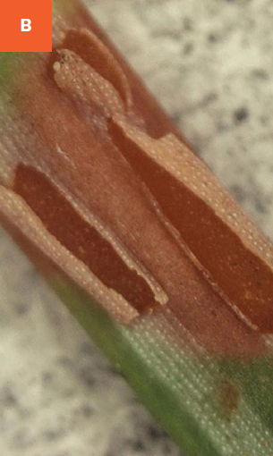
DISEASE CYCLE The fungus overwinters in infected needles. During wet weather in spring, the fungus produces fruiting bodies under the epidermis of spots. Under wet and cool conditions, the epidermis of affected tissues splits lengthwise and masses of spores are released. The spores are dispersed by wind and rain and infect newly emerging needles.
MANAGEMENT Plant resistant varieties in full sun whenever possible. Prune out and discard infected branches during dry weather. Disinfect pruning tools between cuts with 70% ethyl alcohol or a standard household disinfectant spray. Affected trees can be treated with fungicides containing the active ingredient copper hydroxide, copper sulfate, mancozeb, or thiophanate-methyl. Apply fungicides in spring when new needles have grown half their mature length. A second treatment should be applied three to four weeks later when new needles are full grown. Depending on the weather conditions and the product used, a third application might be necessary. Strictly follow instructions on the pesticide labels. Fungicide applications will not cure infected needles but will prevent new infections.
A Epidermis of affected needle tissues splits lengthwise. B Close-up of split epidermis. C Browning needles on Douglas-fir. D Brown spots on infected needles.